11
Mar 2019
Moving ultrasound pictures capture babies' first breaths
Published in General on March 11, 2019
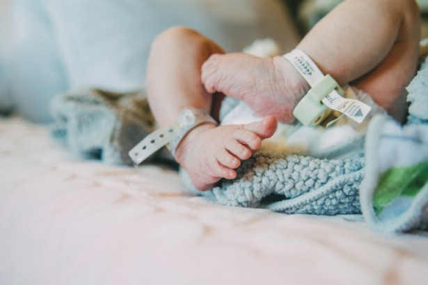
The birth of a baby is something that always never fails to fill parents and healthcare staff with a mixture of trepidation and joy. These early hours in a newborn’s life are critical as the newborn’s lungs first begin to function.
Previously, in the absence of advanced technology, doctors often were unable to provide adequate care and treatment to help newborns born with severe breathing difficulties. Consequently, this led to cases of avoidable brain damage and death.
However, there is hope on the horizon with recent developments as a result of a collaboration between the Royal Women’s Hospital and Monash University. This collaboration enabled doctors to view ultrasound images of a newborn baby’s lungs.
This greater clarity gives doctors a clearer picture which helps them provide life-saving treatment and support if needed. This breakthrough has the potential to save hundreds of lives as doctors are able to immediately diagnose breathing problems in the first few minutes of a newborn’s life. In the past, doctors could only tell if something was wrong after several hours of life which was unfortunately too late for the child.
A deeper study into images captured
By using ultrasound to capture 115 images of the lungs of newborn babies, the researchers at Royal Women’s Hospital in collaboration with Monash University were able to study the developmental patterns of the lungs of newborn babies. This research initiative, known as DOLFIN, also uses lung scans of 28 full-term babies before and after they took their first breaths.
From here, the researchers studied the average time taken for a newborn baby’s lungs to acclimatize to breathing air. Post-deliver, a newborn’s lungs are filled with liquid which are slowly evacuated over time as the lungs aerate and the newborn takes his/her first breaths.
“Within 10 minutes, the lungs of these healthy babies appear to have nearly completed the adjustment needed after birth, although it does take, on average, up to four hours for all the liquid to be removed from the baby’s lungs,” said lead researcher Dr Douglas Blank.
This information is especially critical as doctors can now determine if a newborn child has breathing difficulties based upon ultrasound scans of the lungs. Hence, within 20 minutes, a physician should be able to determine if there are any problems associated with the newborn’s lungs. In this way, corrective action can be very quickly taken to support breathing function which can prevent brain damage or death.
This is especially important in cases of premature birth as premature babies often have underdeveloped breathing functions and lungs. All of this necessitates treatment in a neonatal intensive care unit where special care is taken to provide the newborn child with all the support it needs.
Thanks to the findings of the Royal Women’s Hospital and Monash University researchers, doctors can now quickly determine if a premature baby requires special treatment for breathing difficulties. Given that 50% of premature infants have severe breathing problems which require intensive treatment, the time saved will no doubt have a momentous life-saving effect for premature babies everywhere.
New breakthroughs
Headed by Dr Blank, a new research project called DOLFIN Jr is currently being undertaken to determine whether imaging of the lungs in the first few minutes of life would be able to predict which premature infants required intensive breathing support. This has the benefit of saving the time of healthcare staff while also helping them prioritize which infants would require their help.
To treat breathing difficulties in premature infants, a drug called Surfactant is used. This drug is administered via inserting a breathing tube into the infant’s airway and placing the child on a ventilator. One of the risks associated with this procedure is the likelihood of serious lung damage which is why this treatment is reserved for only the most severe cases.
For premature infants without severe breathing problems, CPAP breathing assistance is used to minimize the likelihood of lung and brain damage. However given that window to decide which treatment should be used is extremely small, lung ultrasound scans may be the best way to save the lives of infants.

